
-
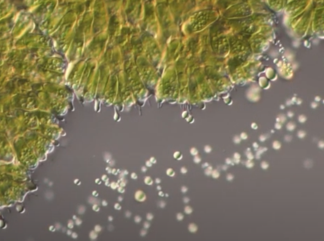
Botryococcus brauni
oil droplets secreted from colony
-
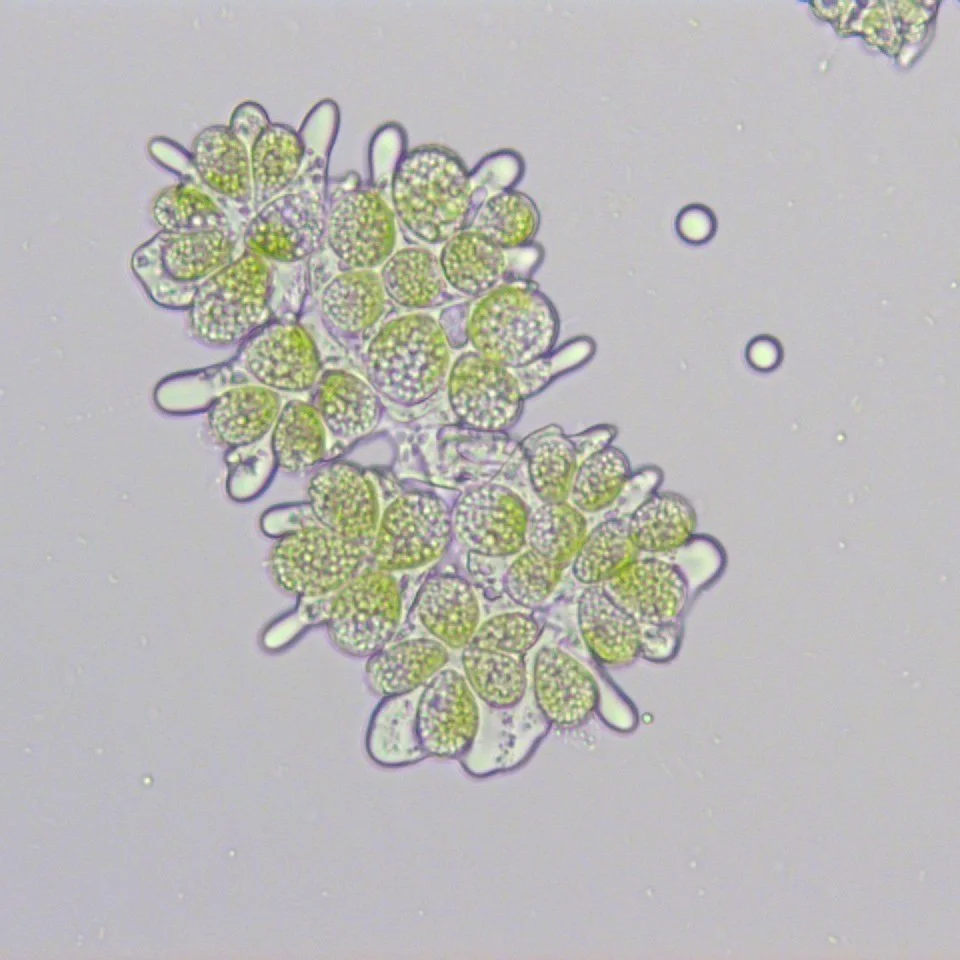
Botryococcus brauni Showa strain
Fat with hydrocarbons and blebbing oil droplets
-
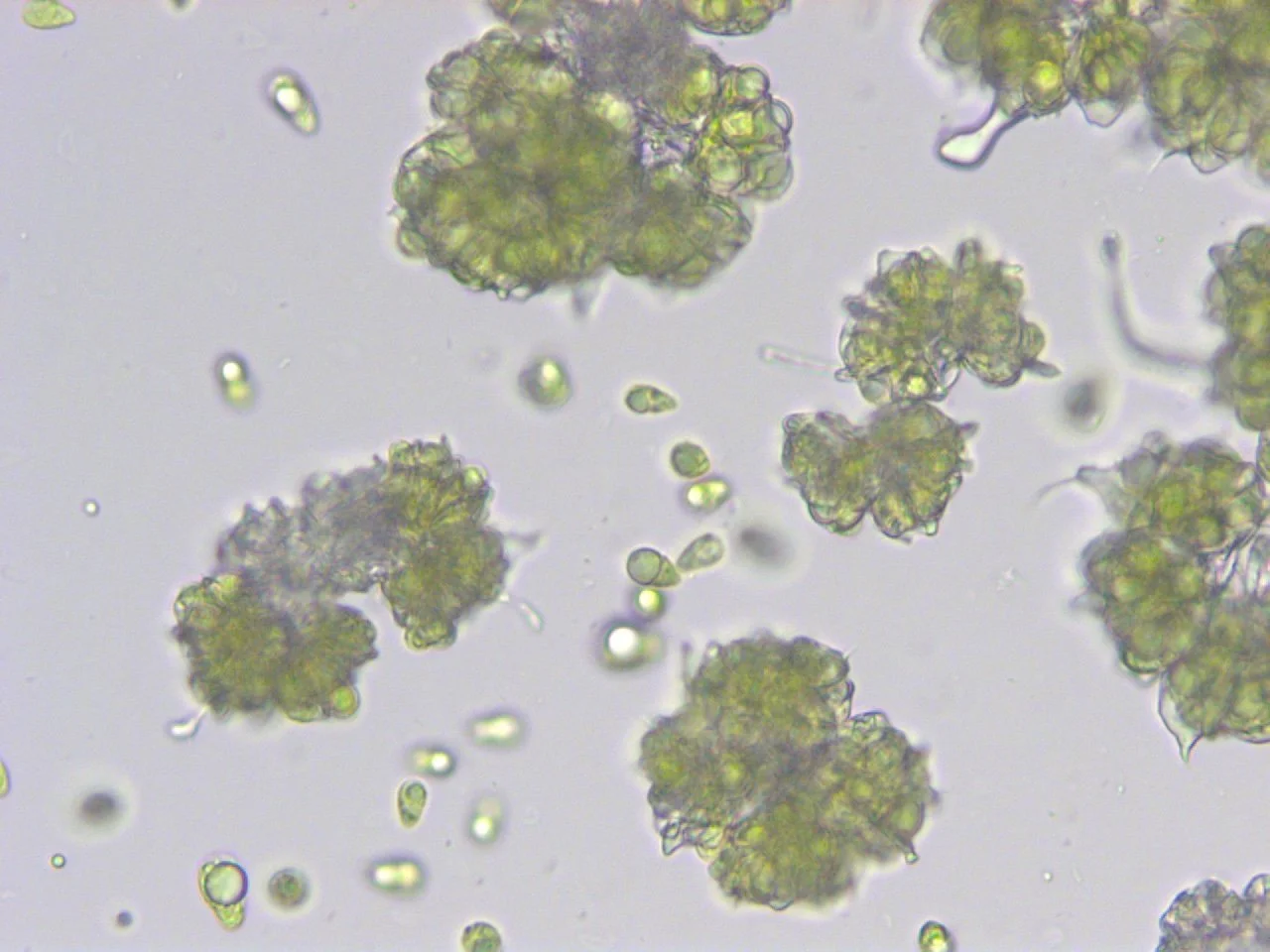
Botryococcus braunii Showa strain
Ejecting single cells bloated with oil from colonies. Why are they doing this? Stressed? Making new daughter colonies?
-
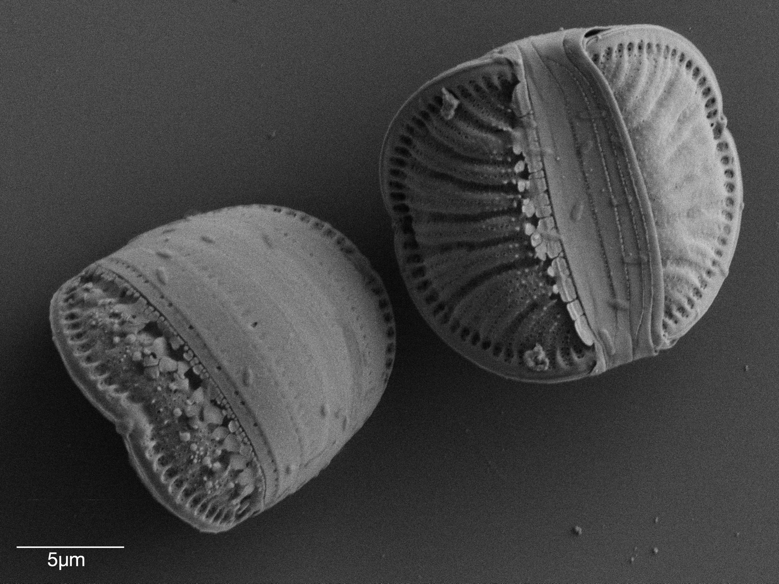
Epithemia
Scanning electron microscopy, looking like a mandarin orange slice
-
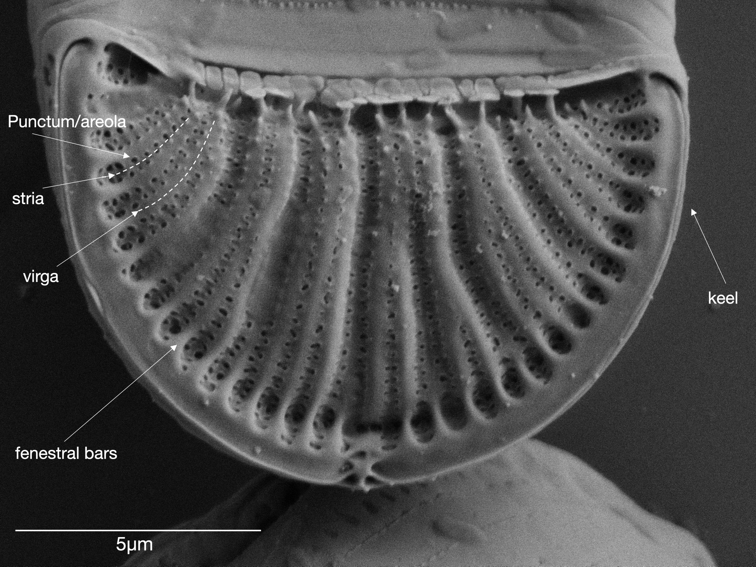
Epithemia
Scanning electron microscope. Thanks, Sarah, for figuring out what’s what!
-
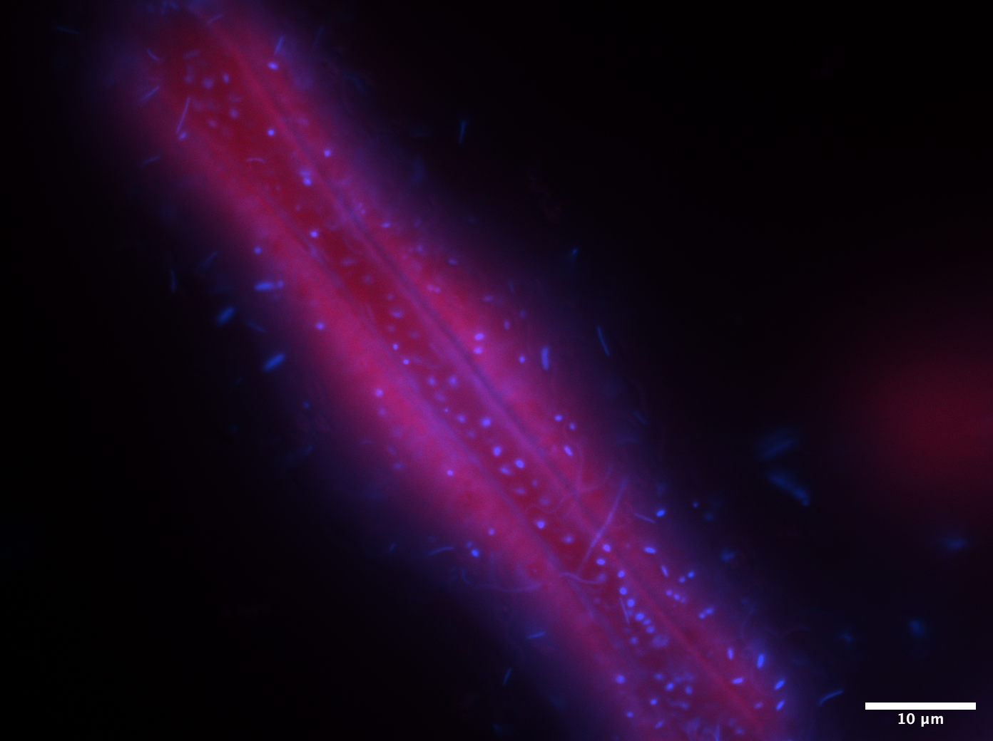
Epithemia
Composite fluorescent image showing chlorophyll auto-fluorescence and DAPI staining. When focused on the surface of the diatom cell, light refracts through the silica shell and results in a DAPI glow and shadows created by thicker features in the silica shell appear.
-
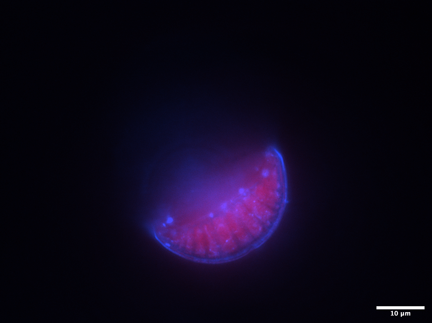
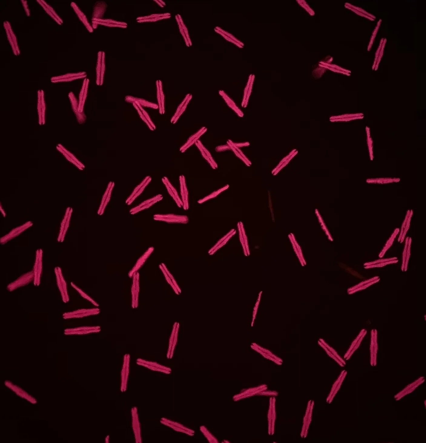
Rhopalodia gibba
Composite fluorescent image showing chlorophyll auto-fluorescence and DAPI staining. The focal plain is at the bottom face of the cell, revealing the bacterial community associating with the diatom.
-
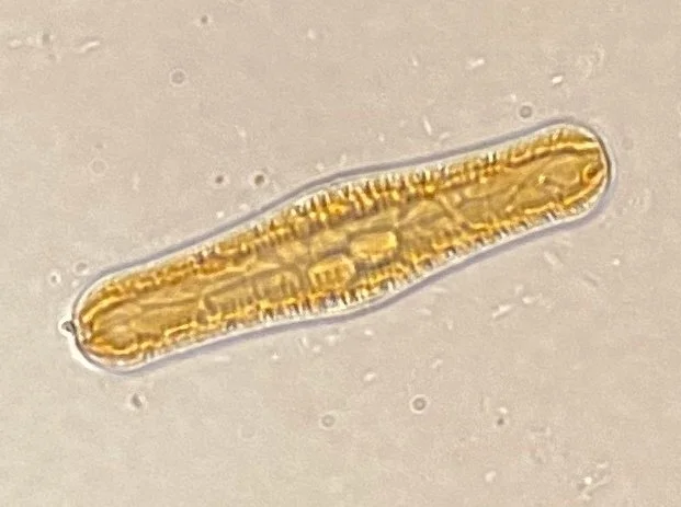
Rhopalodia gibba
Light microscope image of rhopalodia gibba. Two ‘Spheroid body’ cyanobacterial endosymbionts can be seen in the center of the cell.
-
Rhopalodia gibba
Fluorescent image of a culture of rhopalodia gibba. The fluorescent property of chlorophyll makes cells appear red.
-
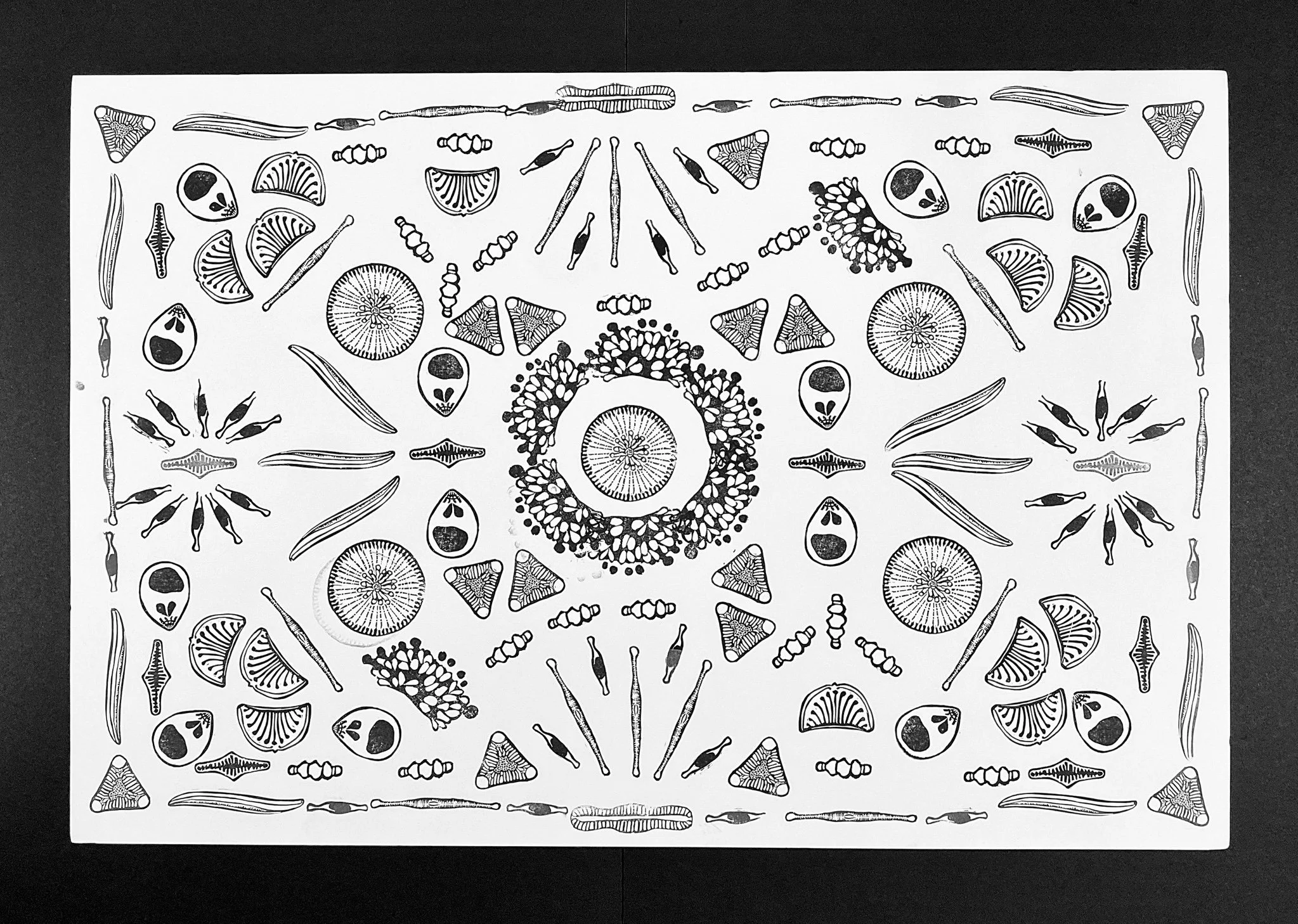
Algae stamp art
Yeh lab happy hour art with stamps
-
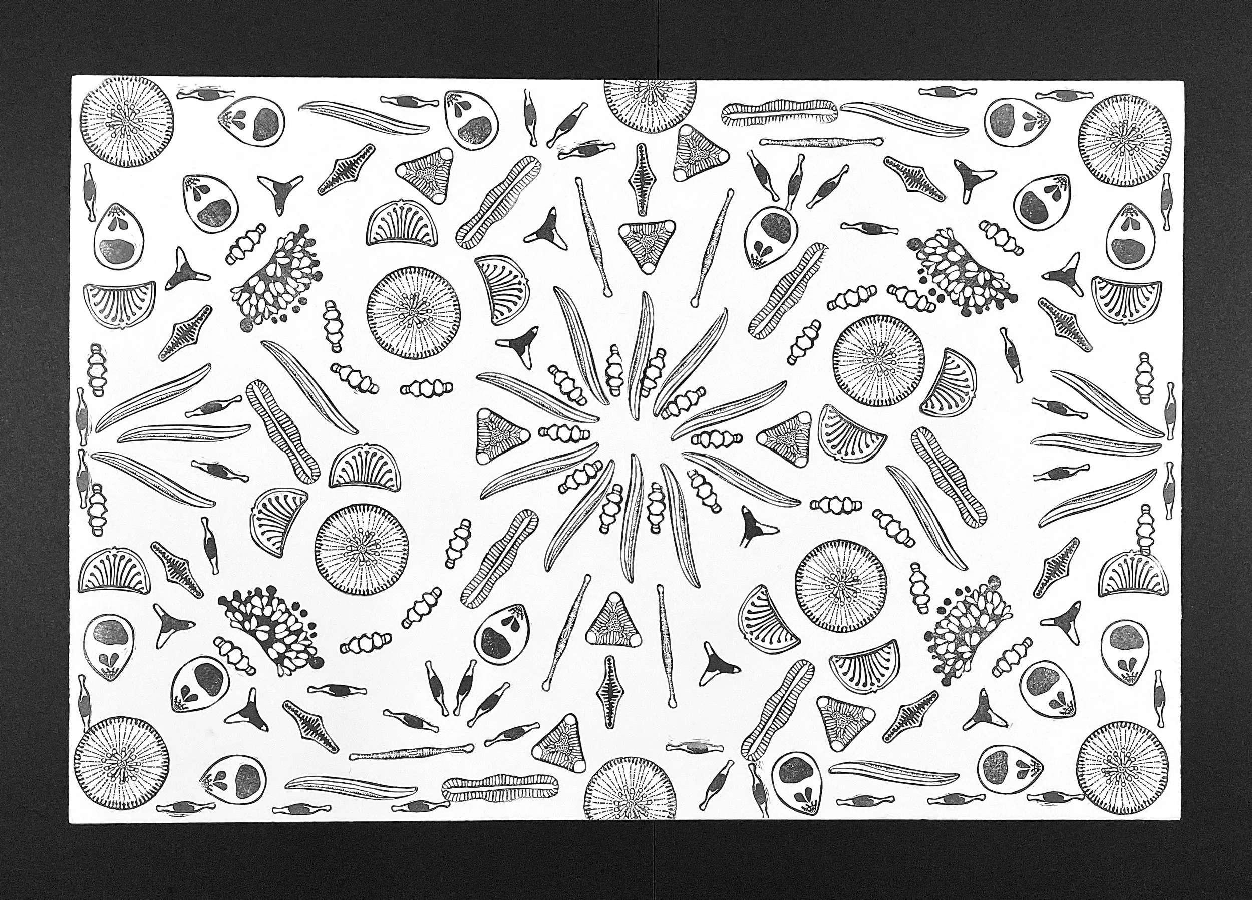
Algae stamp art
-
New List Item
Description goes here